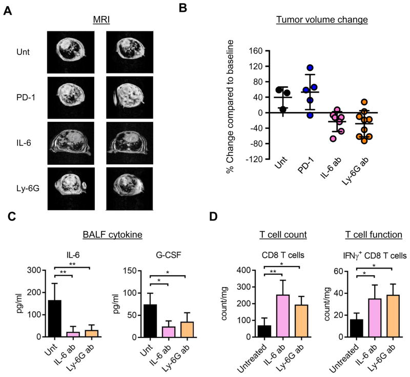
Figure 4. IL-17 high tumors are resistant to PD-1 immune checkpoint blockade but sensitive to IL-6 blockade or neutrophil depletion.
A) Left. Representative Magnetic resonance imaging (MRI) images and Right: Quantification of MR images of mice untreated or treated with PD-1, IL-6 or Ly-6G blocking antibodies for 2 weeks. B) Quantification of MRI for treatments in A. Each data point represents a different mouse. C) Ki67 IHC on the lung tissue from IL-17:Kras mice either untreated or treated with IL-6 antibody, and quantification of IHC. p=0.02 for Ki67 counts between untreated and IL-6 antibody treated IL-17:Kras tumors. Each data point is from a different mouse lung tissue. D) Levels of IL-6 and G-CSF in the Bronchoalveolar lavage fluid of IL-17:Kras mice untreated (n=4) or treated with IL-6 (n=5) or Ly-6G (n=5) antibodies for two weeks. E) CD8 T cell counts (left) and intracellular interferon gamma positive CD8 T cell counts per mg of lung for IL-17:Kras mice either untreated or treated with IL-6 or Ly-6G antibodies. Untreated (n=4), treated with anti-IL-6 treated:IL-6 ab (n=5) and anti-Ly-6G treated:Ly-6G ab (n=5).

