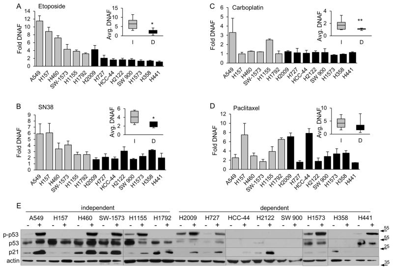
Figure 2. KRAS dependent NSCLC cells show reduced sensitivity to DNA damaging agents.
(A–D), K-Ras independent (gray bars) or K-Ras dependent (black bars) cells were treated for 24 hours with 50 μM etoposide (A), 10 μM SN38 (B), or for 48 hours with 20 μg/ml carboplatin (C), or 10 nM pacilitaxel (D), and DNA fragmentation was assayed as described in Materials and Methods. The range of DNA fragmentation values from K-Ras independent (I, gray bars) and dependent (D, black bars) cell lines is shown in the inset for each graph with the black line indicating the median. Data shown is the average of 3 or more independent experiments; * = p <0.05, ** p <0.09. (E) NSCLC cells were treated with 50 μM etoposide for 24 hours. Phosphorylation of p53 at Ser15 and expression of p21 was assayed by immunoblot as described in Materials and Methods. Blots were stripped and probed for actin and a total p53 antibody which detects WT p53 and some forms of mutant p53.

