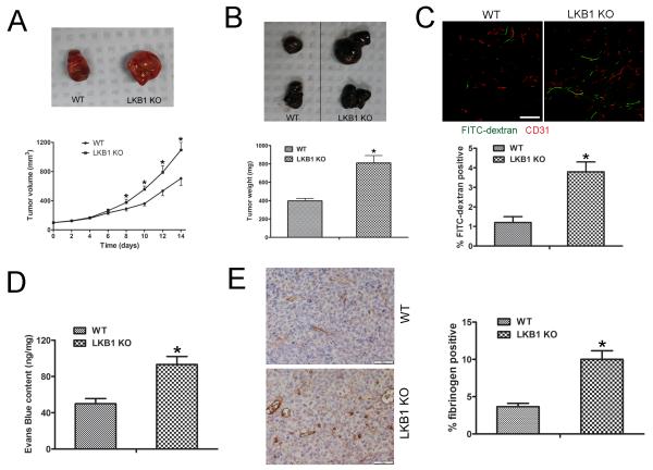
Figure 2. Tumors implanted into LKB1endo−/− mice show increased growth and microvascular permeability.
A, LLC cells (106) were implanted subcutaneously into the lower backs of 6- to 8-week-old male WT or LKB1endo−/− mice. Tumors were allowed to grow for 14 days. P< 0.05, compared with WT (n = 8). Representative images of tumors. B, B16F10 melanoma cells (106) were implanted subcutaneously into the backs of WT or LKB1endo−/− mice for 14 days. Representative images are shown and the weight of tumors was quantified. P < 0.05, compared with WT (n = 8). C, LLC tumor-bearing mice were injected with 70-kDa FITC-conjugated dextran to visualize vessel permeability, then tumor sections were immunostained for CD31 (red); FITC-conjugated dextran appears in green. Bar = 50 μm. Blood vessel permeability was analyzed by area of extravasated dextran. *, P < 0.05, compared with WT (n = 5). D, Vascular permeability in implanted LLC tumors measured by Evans blue extravasation (30 mg/kg intravenously, 30 min) as an index of albumin leakage. *, P < 0.05, compared with WT (n = 5). E, Frozen sections (5 μm) of LLC tumors were stained with fibrinogen and quantified. Bar = 50 μm. *, P < 0.05, compared with WT (n = 5). Data are mean ± SE.

