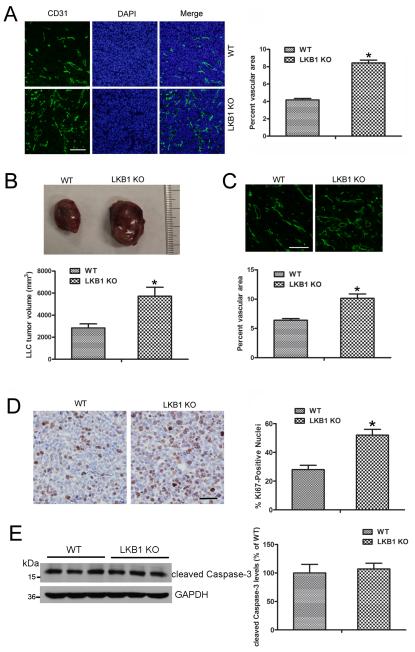
Figure 3. Tumors implanted into LKB1endo−/− mice exhibited increased angiogenesis.
A, Frozen sections of 2-week LLC tumors were stained with CD31, and the vascular area in LLC tumors was quantified. Bar = 100 μm. *, P< 0.05, compared with WT (n = 6). B, LLC cells were implanted into WT or LKB1endo−/− mice. After 28 days, tumors were harvested and quantified. *, P< 0.05, compared with WT (n = 8). C, Frozen sections of 4-week LLC tumors were stained with CD31 and the vascular area was quantified. Bar = 100 μm. *, P< 0.05, compared with WT (n = 6). D, Representative Ki-67 immunohistochemical images. Bar = 50 μm. Ki-67–positive signals in LLC tumors were quantified. *, P < 0.05, compared with WT (n = 5). E, Western blot analysis of cleaved Caspase-3 level in LLC tumors (n=6). Data are mean ± SE.

