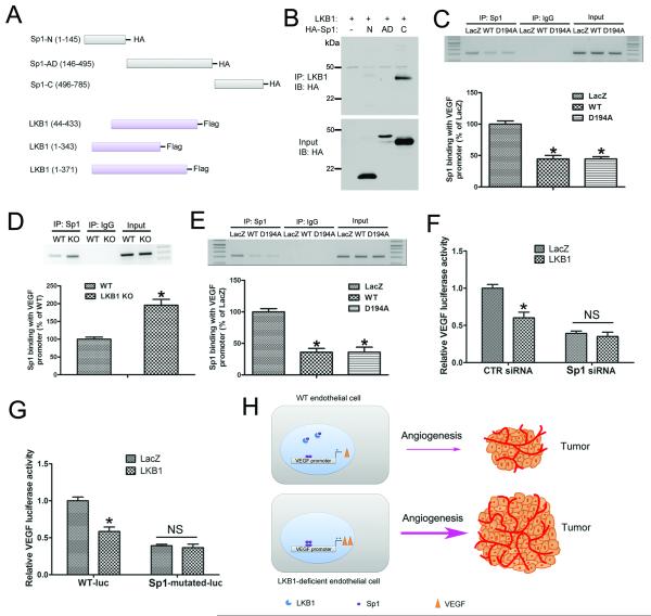
Figure 7. LKB1 binding with Sp1 inhibits VEGF expression.
A, A schematic diagram showing the construction of truncated LKB1 and Sp1 plasmids. B, HEK293T cells were transfected with WT LKB1 and HA-tagged truncated Sp1 plasmids, and cell lysates were immunoprecipitated with LKB1 antibody, then underwent western blot analysis with anti-HA antibody. C, A549 cells were transfected with LacZ, WT or D194A LKB1 plasmid, then underwent DNA ChIP assay to detect the ability of Sp1 to bind to the VEGF promoter. *, P< 0.05, compared with LacZ (n = 5). D, WT and LKB1endo−/− ECs were used in ChIP assay to detect the ability of Sp1 to bind to the VEGF promoter. *, P< 0.05, compared with WT (n = 5). E, LKB1endo−/− ECs were transfected with LacZ, WT or D194A, then underwent DNA ChIP assay. *, P< 0.05, compared with LacZ (n = 5). F, Luciferase activity assay of A549 cells transfected with CTR siRNA or Sp1 siRNA for 48 hr, then with LacZ or LKB1, together with VEGF promoter-luciferase plasmids for 24 hr. *, P< 0.05, compared with LacZ (n = 5). G, Luciferase activity assay of A549 cells transfected with LacZ or LKB1, together with WT or Sp1-mutated VEGF promoter-luciferase plasmids, for 24 hr. *, P< 0.05, compared with LacZ (n = 5). H, A diagram showing the mechanism by which LKB1 regulates endothelial angiogenesis and tumor growth via VEGF. Data are mean ± SE.

