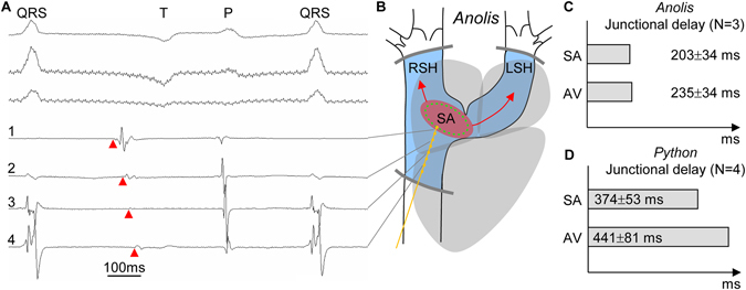
Figure 3.

The sinuatrial junctional area harbors the dominant pacemaker, has a delay, and the sinus venosus is activated retrograde in both Anolis and Python. (A) Original traces of three-lead surface ECG (upper part of the panel) and local recordings from 4 bi-polar electrodes in Anolis equestrie (traces 1 to 4 in lower part of the panel). Electrode 1 is in the sinuatrial junctional area, whereas electrode 4 is the furthest into the posterior sinus horn. Deflections in all 4 electrodes can be seen to align with the P- and QRS-waves of the ECGs and these deflections are considered remote activity. The deflections indicated by red arrowheads occur prior to the deflections aligned with the P-wave and are therefore considered local electrograms of the sinus venosus. The earliest deflection is in electrode 1 and the latest in electrode 4, thereby showing dominant pacemaker activity in the sinuatrial junctional area and retrograde activation of the posterior sinus horn. In electrode 1, the interval between the early deflection (red arrowhead) and the deflection aligned with the P-wave constitutes the substantial sinu-atrial delay. (B) All sinus horns are activated retrograde from the sinu-atrial junctional area and the activation front stops at the pericardial reflection. (C,D) Within species, the sinu-atrial (SA) and atrioventricular (AV) delays are of similar duration. LSH, left sinus horn; RSH, right sinus horn.
