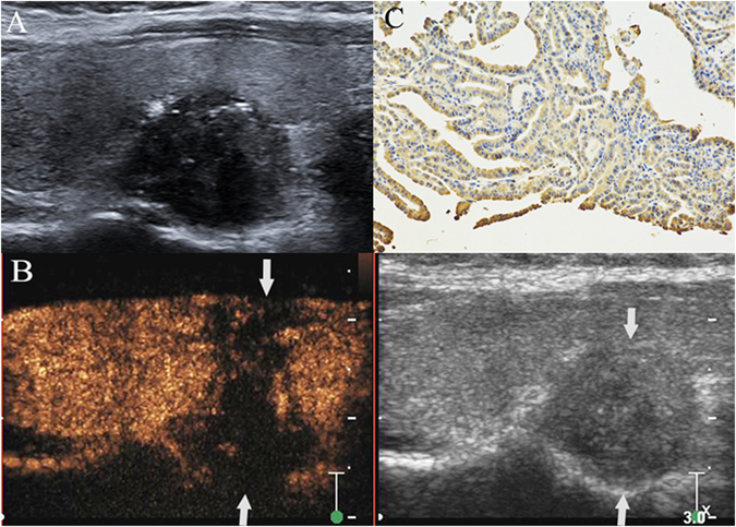
Figure 1.

PTC with cervical LNM. A hypoechoic nodule with ill-defined margin, irregular shape and some microcalcifications was found at the lower pole of the right thyroid lobe in a 34- year– old female patient (A). The thyroid focal posterior capsular was bulged by nodule. The size of nodule was 1.6 cm × 1.5 cm × 1.0 cm. CEUS showed that a heterogeneous low-enhancement in nodule (left image) and its area (1.8 cm × 1.6 cm × 1.2 cm) was larger than the nodule region displayed on grey-scale image (right image) (arrow). Both the anterior and posterior thyroid capsular were broken and invaded by the nodule (arrows) (B). Intense expression (+++) (arrow) of matrix metalloproteinase-9 was found in the cytoplasm of the malignant cells of a PTC sample, as shown by the brown staining (magnification, ×200) (C). Surgical results: PTC with ipsilateral region III-V and central compartment region LNM.
