
Figure 1.
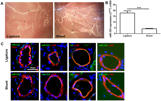
Expression of miR-352 in collateral vessels of the ligature-only side (LO) and the AV-shunt side (AVS). (A) Collateral vessels that were infused with liquid latex in the superficial adductor muscles from both the LO and AVS. An increased number of collaterals was evident in the AVS (indicated by arrows); original magnification: 10×. (B) Quantitative analysis of miR-352 expression by stem-loop qPCR in LO and AVS collateral vessels from each animal (***P < 0.001, n = 5). (C) In situ hybridization from miR-352 in collateral vessels of the LO and AVS. Green fluorescence and red fluorescence for CD31 and α-SM-actin, respectively. The U6 probe was used as a positive control, and miR-159 served as a negative control because this miRNA is not expressed in mammalian tissue. Notably, miR-352 was present mainly in endothelial and smooth muscle cells of the collateral vessels, and its expression was decreased in collateral vessels of the AVS as compared with those in the LO.