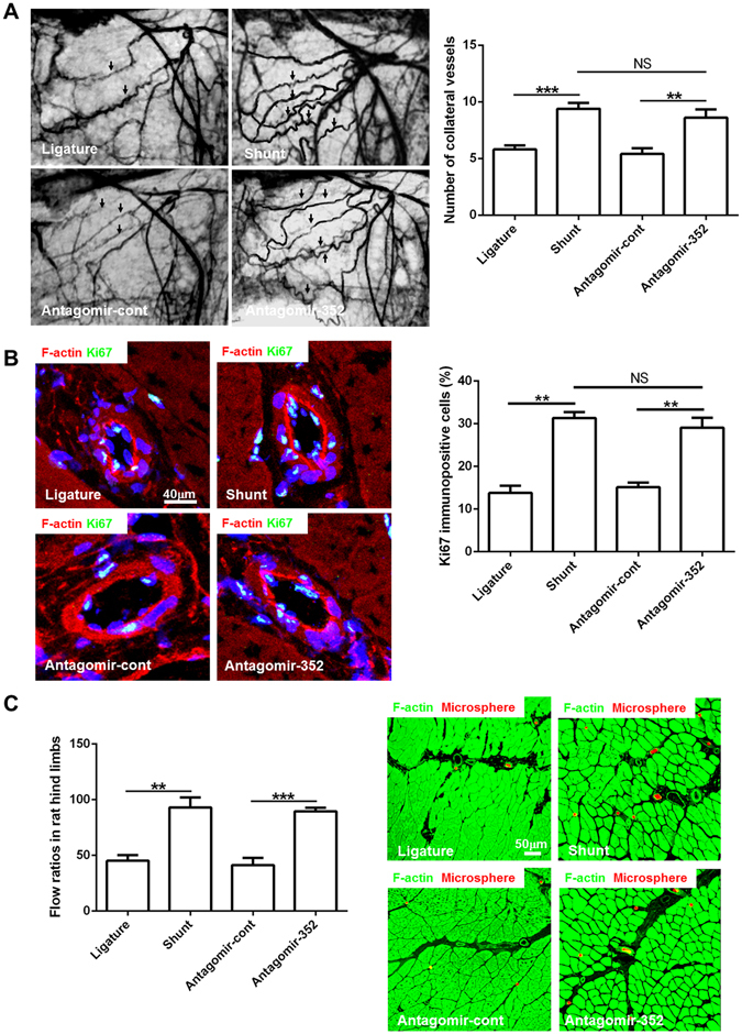
Figure 3.

Inhibition of miR-352 enhances arteriogenesis and promotes flow restoration in rat hind limbs. Ligature: the ligature-only side; Shunt: the AV shunt side; antagomir-352: rats infused with antagomirs targeting miR-352 through an osmotic mini pump connected to the proximal region of the occluded femoral artery; and antagomir-cont: control antagomir. (A) Postmortem angiograms of rat hind limbs on day 7 after infusion. Antagomir-352 treatment resulted in an increase in the number of collateral vessels (arrows) as compared to antagomir-cont treatment. This increase is similar to the number of collateral vessels observed following shunt treatment. (***P < 0.001, **P < 0.01, n = 5). (B) Cell proliferation was detected by Ki67 immunostaining in collateral vessels from different groups. Green fluorescence for Ki67, red for F-actin, blue for nuclei. Notably, Ki67-positive cells were more numerous in collaterals treated with antagomir-352 than in the control side, which is similar to what is observed following shunt treatment (**P < 0.01, n = 5). (C) Flow ratio of rat hind limbs at day 7 after antagomir-352 infusion and fluorescence microspheres administration in adductor muscles. Antagomir-352 treatment resulted in a significant increase in collateral flow, compared to antagomir-cont treatment. This increase is similar to that observed following shunt treatment (**P < 0.01, ***P < 0.001, n = 5). Red fluorescence for the fluorescence microspheres, and green for F-actin. The number of fluorescence microspheres was higher in the antagomir-352-treated and AV shunt-side muscles.
