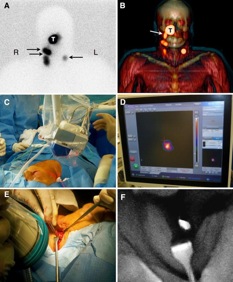
Fig. 2.
Combined preoperative lymphatic mapping and intraoperative radio- and fluorescence-guided sentinel node biopsy. a Early static anterior preoperative lymphoscintigram 10 min after infraorbital peritumoral injection of ICG–99mTc-nanocolloid showing the injection site (T) with lymphatic drainage to two sentinel nodes in the neck on the right (R) side and a third one on the left (L) side (arrows). b 3D SPECT/CT image 2 h post-injection providing additional anatomical information with visualisation of a lymphatic duct (arrow) originating from the injection site (T). c, d Intraoperatively, the radioactive component of the hybrid tracer in the left sentinel node is visualised using a portable gamma camera, and its laser pointer guides placement of the incision. e, f A near-infrared fluorescence camera is used to visualise the fluorescent component of the hybrid tracer in the same (non-blue) sentinel node
(Figure reproduced with permission from [40])

