Figure 1.
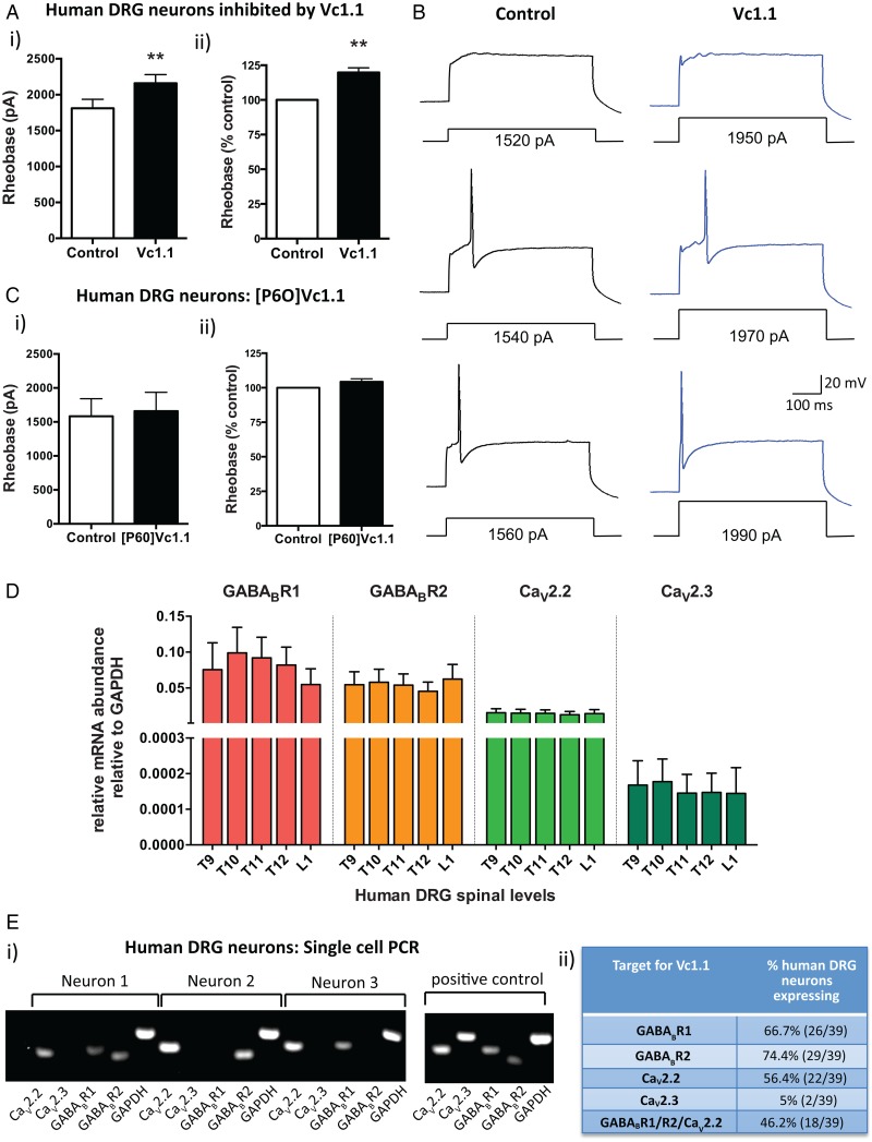
α-Conotoxin Vc1.1 inhibits human dorsal root ganglion (DRG) neurons. (A) (i) Group data showing that Vc1.1 (1000 nM) significantly increases the rheobase of a subpopulation (40%) of human DRG neurons, indicating Vc1.1 inhibits neuronal excitability and more current is required to initiate an action potential (**p<0.001, n=10, paired t-test). (ii) In this population of neurons, Vc1.1 increased the rheobase by 20% compared with baseline response, meaning the neurons are less excitable (**p<0.001). (B) Representative examples of human DRG neuronal responses in the absence (control solutions) and in the presence of Vc1.1. Note in this example more current is required to fire an action potential from a human DRG neuron in the presence Vc1.1 (1970 pA) relative to control (1540 pA). (C) [P6O]Vc1.1 (1000 nM), a synthetic analogue of Vc1.1 that does not act at γ-aminobutyric acid receptor B (GABABR), did not affect human DRG neuronal excitability when expressed as either i) rheobase or ii) % of rheobase (p>0.05, n=10, paired t-test) indicating Vc1.1 induces its inhibitory effect via the GABABR. (D) Group data of quantitative-reverse-transcription-PCR analysis from thoracolumbar (T9, T10, T11, T12, L1) DRG from four human adult donors indicating expression of the GABABR subunits R1, R2 and the voltage-gated calcium channels CaV2.2 and CaV2.3 in human DRG. (E) (i) Examples of gel electrophoresis following single-cell-PCR analysis from individual human DRG neurons. (ii) Combined analysis of expression and coexpression of GABABR and CaV channels in 39 human DRG neurons. Of human DRG neurons, 46.2% (18/39) coexpress GABABR and CaV2.2, the minimum components required for Vc1.1-induced inhibition. Combined these studies indicate Vc1.1 inhibits human DRG neurons via a GABABR-mediated mechanism. GAPDH, glyceraldehyde-3-phosphate dehydrogenase.

