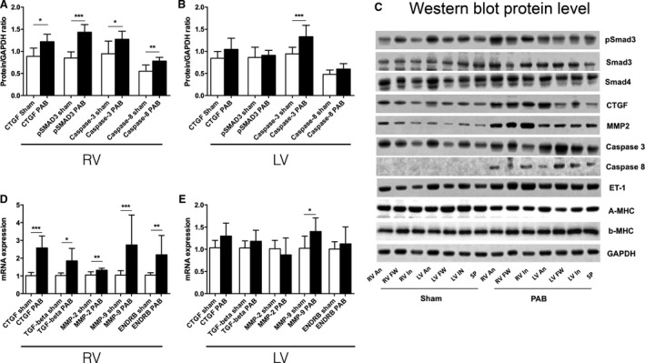
Figure 5.

(A and B) Regional right and left ventricular hinge‐point region protein levels (Western blot) in PAB rabbits. Phosphorylated SMAD3 and CTGF, which acts downstream of TGF β1, is elevated at the right ventricular hinge‐point regions. The apoptosis related enzymes caspase‐3 and ‐8 in the right ventricular hinge‐point region and caspase‐3 at the left ventricular hinge‐point region are increased compared with shams (Sham: n = 5 and PAB: n = 3). (C) Western blot gel examples. (D and E) Real time PCR results from the right and left ventricular hinge‐point regions. Gene expression of profibrotic signaling and extracellular matrix remodeling including CTGF, MMP‐2, MMP‐9, and endothelin receptor B (ENDRB) are up‐regulated in the RV hinge point region in PAB rabbits compared with shams, (Sham: n = 6 and PAB n = 3). Data are presented as mean (SD). *P < 0.05, **P < 0.01, ***P < 0.001 RV, right ventricle and LV, left ventricle.
