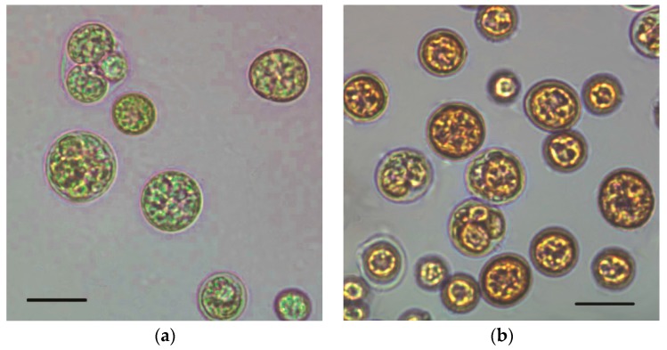
Figure 1.
Microscopic images of C. zofingiensis cells during mixotrophic growth. (a) Initial stage at 0 h; (b) Final stage at 288 h; Bars: 10 μm. The mixotrophic growth experiment was performed in modified basal medium with 30 g/L of glucose and 0.6 g/L of NaNO3 with a high C/N ratio of 200. A light intensity at 80 μmol photon m−2 s−1 was employed for astaxanthin accumulation after inoculation of seed culture grown mixotrophically under 10 μmol photon m−2 s−1.

