
Figure 3. Some Pax2-deficient cap mesenchyme cells can persist throughout kidney development.
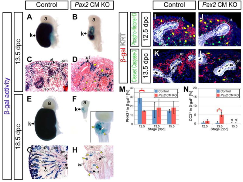
(A–H) Kidneys from Six2-eGFPCretg/+; Pax2flox/+; R26RlacZ/+ control mice (Control) and Six2-eGFPCretg/+; Pax2flox/del; R26RlacZ/+ cap mesenchyme-specific Pax2 mutant mice (Pax2 CM KO). X-gal staining of whole-mount tissues (A,B,E,F) and sections counter-stained with eosin (C,D,G,H) at 13.5 dpc (A–D) and 18.5 dpc (E–H). Yellow arrows and yellow arrowheads in D indicate β-gal+ cap mesenchyme-derived cells around the ureteric tip with less columnar cell shape and spreading away from the ureteric tip, respectively. The inset in F shows a high magnification of a hypoplastic kidney in Pax2 CM mutants. Yellow arrowheads and white arrow in F and H indicate β-gal+ cap mesenchyme-derived cells in the interstitium and in a few rare nephrons, respectively. (I–L) Confocal immunofluorescence of the nephrogenic zone of kidneys from Control mice (I,K) and Pax2 CM KO mice (J,L) with β-gal (red) and cytokeratin (KRT; ureteric tip, white) staining. (I,J) Phospho-histone H3 (green) staining at 12.5 dpc. Yellow arrows indicate β-gal+ phospho-histone H3+ cells. (K,L) Cleaved caspase-3 staining at 13.5 dpc. Yellow arrows in M indicate β-gal+ cleaved caspase-3+ cells. (M and N) Quantification of phospho-histone H3+ (PHH3+) (M) and cleaved caspase-3+ (CC3+) (N) cells in β-gal+ cap mesenchyme-derived cells. Data are represented as mean ± SD. Asterisks indicate p<0.05 with unpaired t-test. n.d., not detected. Abbreviations as in Figure 2.