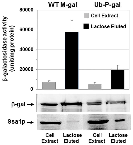
Fig. 3. Ssa1p was preferentially associated with the rapidly degraded Ub-P-gal.

Wild-type cells expressing M-gal or Ub-P-gal were grown at 28°C in the presence of 2% galactose for 12–16 h. 3–4 mg of cell proteins were prepared and applied to a 1-ml column of p-aminobenzyl-1-thio-β-galactoside crosslinked to agarose beads at 4°C. The bound β-gal were eluted with lactose and the presence of Ssa1p or β-gal fusion proteins was determined by Western-blot with anti-human Hsp70 or anti-β-gal antibodies and also the β-gal activity was also measured.
