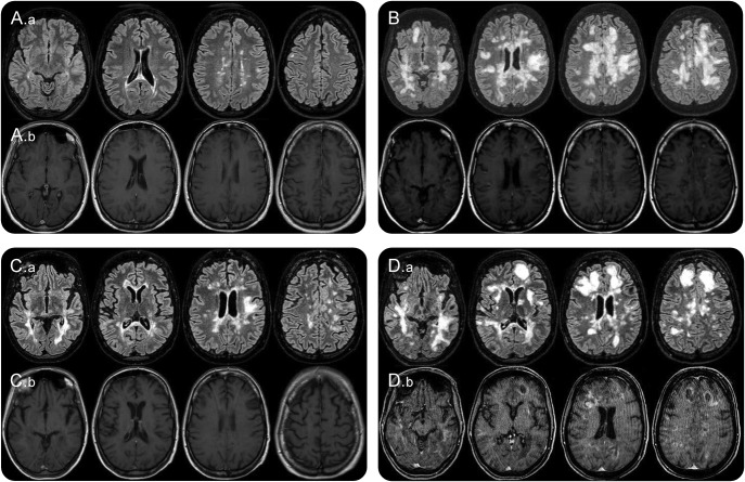
Figure. Patient 1 MRI scans.
(A) Brain MRI performed on October 12, 2012. (A.a) FLAIR (FLuid Attenuated Inversion Recovery) sequence. (A.b) T1 gadolinium enhanced sequence. Routine MRI scan during fingolimod therapy shows some periventricular juxtacortical FLAIR white-matter hyperintensities with no gadolinium enhanced lesions. (B) Brain MRI performed on August 29, 2013. In the FLAIR sequence diffuse and confluent white-matter hyperintensities are found in both hemispheres in the periventricular and subcortical white matter, with the involvement of the “U” fibers. Many lesions show gadolinium enhancement. (C) Brain MRI performed on October 17, 2013. One month after cyclophosphamide IV administration. FLAIR white-matter hyperintensities are significantly reduced (C.a) and no lesion display gadolinium enhancement (C.b) compared with the MRI performed on 29th of August (B). (D) Brain MRI performed on January 3, 2014. A remarkable increase in the number and size of white-matter hyperintensities can be observed (FLAIR sequence, D.a) compared with the scan of 17th of October. New diffuse and rim gadolinium enhancing lesions are present (D.b).

