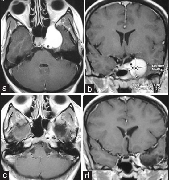
Figure 1.

Preoperative contrast magnetic resonance imaging of brain; (a) axial and (b) coronal images showing highly contrast enhancing tumor (haemangioma) in left cavernous sinus, (c and d) postoperative contrast magnetic resonance imaging in axial and coronal images respectively showing very small residual tumor around the posterior cavernous ICA
