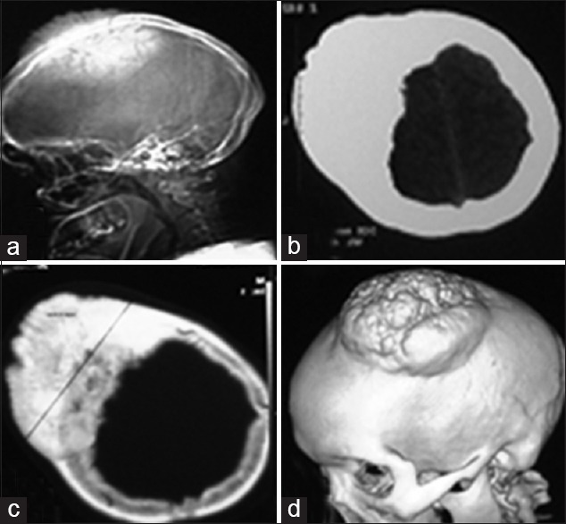
Figure 1.

(a) Plain lateral radiograph showing osteoma in the frontoparietal region. (b) Non contrast computed tomography (CT) scan section showing hyperdense area on the right frontoparietal bone. (c) CT scan bone window showing excessive bone hyperthropy. (d) Three dimensional reconstruction showing giant osteoma on left parietal region
