Figure 3.

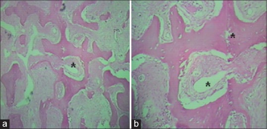
(a) Histopathologic image of osteoma (H and E, ×10) showing dense lamellae with organized haversian canals (*) and (b) the intratrabecular stroma contains osteoblasts, fibroblasts, and giant cells, with no hematopoietic cells

(a) Histopathologic image of osteoma (H and E, ×10) showing dense lamellae with organized haversian canals (*) and (b) the intratrabecular stroma contains osteoblasts, fibroblasts, and giant cells, with no hematopoietic cells