
Figure 1.
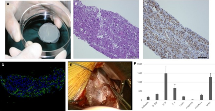
Characterization of implanted autologous skeletal stem‐cell sheets. A, The appearance of cell sheets. B, Hematoxylin and eosin staining. C, Desmin staining. D, Fibronectin staining. E, Cell sheets were implanted to LV free wall via left thoracotomy. F, In vitro study; various cytokines were detected in supernatant of cell sheet. G‐CSF indicates granulocyte colony‐stimulating factor; HGF, hepatocyte growth factor; IL‐8, interleukin‐8; PDGF‐BB, platelet‐derived growth factor‐BB; PECAM‐1, platelet endothelial cell adhesion molecule; VEGF, vascular endothelial growth factor.