Figure 4.

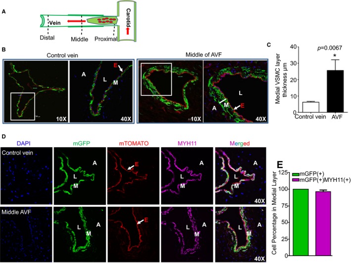
Mature vascular smooth muscle cells (VSMCs) contribute to arteriovenous fistula (AVF) maturation. A, Schematic illustration of the AVF regions used for immunofluorescence staining and histological examination in Myh11‐Cre/ERT2‐mTmG reporter mice at 4 weeks after AVF surgery. B, Representative images for mT‐ or mG‐labeled vessel wall of the control jugular vein branch and the middle AVF region in tamoxifen‐induced Myh11‐Cre/ERT2‐mTmG mice at 4 weeks after AVF surgery. C, Quantitative analysis of the thickness of the medial VSMC layer in the control jugular vein branch and the middle AVF region as described in (B) by software provided by the confocal microscope machine (n=4). There is medial VSMC layer thickening compared with control unligated vein branch at this time point. D, Immunofluorescence staining for MYH11in samples, as described in (A). Representative images from 4 different animals are shown. E, Quantitative analysis for the percentage of MYH11 (+) green fluorescent protein (+) cells in the medial VSMC layer of the middle AVF region by ImageJ software (n=4). The thickened medial layer is comprised of contractile VSMCs derived from previously differentiated mature VSMCs. A indicates adventitia; DAPI, 4′,6‐diamidino‐2‐phenylindole; E, endothelium; L, lumen; M, medial smooth muscle layer; mGFP, membrane green fluorescent protein.
