Figure 5.

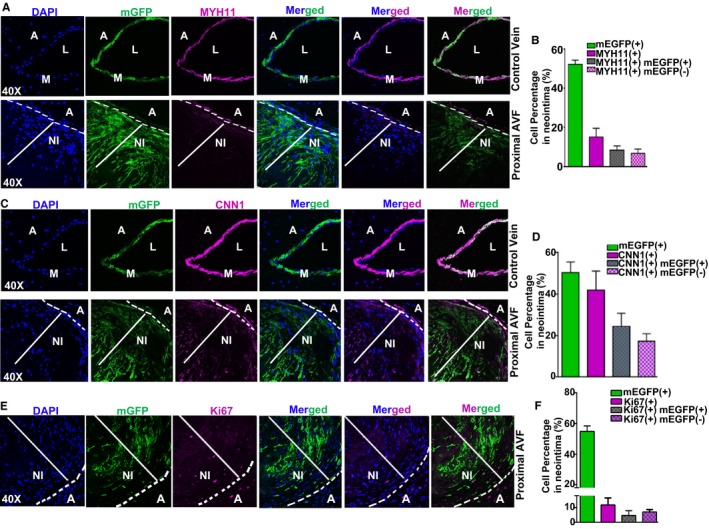
Mature vascular smooth muscle cells (VSMCs) contribute to neointima (NI) formation in arteriovenous fistula (AVF). Confocal microscopy for membrane green fluorescent protein (mGFP) and immunofluorescence staining for MYH11 (A) and CNN1 (C) in the control unligated vein and proximal AVF region, and Ki67 (E) in the proximal AVF region of Myh11‐Cre/ERT2‐mTmG mice at 4 weeks after AVF surgery. Quantitation of the percentage for each indicated cell category was analyzed using ImageJ and results are shown in B, D, and F, respectively (n=4). Extensive NI formation frequently occurs at the proximal region to the anastomosis site. A large portion of NI cells are GFP+ and with decreased levels of contractile proteins including MYH11 and CNN1. A indicates adventitia; DAPI, 4′,6‐diamidino‐2‐phenylindole; L, lumen; M, medial smooth muscle layer.
