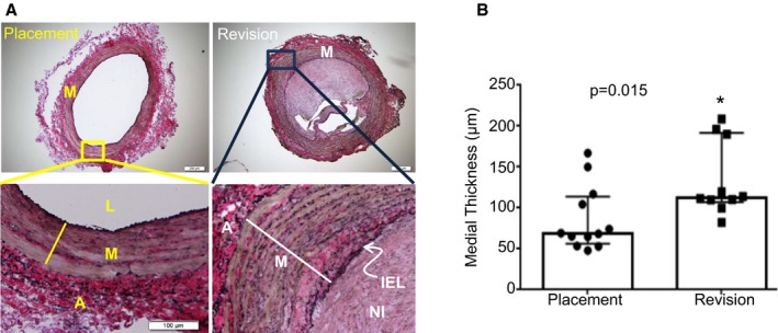
Figure 6.

Morphometric analysis of human venous samples. A, Representative images of Verhoeff‐van Gieson stainings for cross sections of tissue samples obtained from patients undergoing arteriovenous fistula (AVF) placement or revision. The Verhoeff‐van Gieson staining marked elastin fibers in black, collagen in red‐pink, and the cell‐rich medial layer in brown. The bottom images are a zoomed‐in version of the upper panel. B, The medial‐intimal thickness of placement and revision vessels was measured, as described in the Materials and Methods section. There was a statistically significant increase in the thickness of the medial layer in the revision AVF samples compared with the placement venous vessels. The bottom and top boundaries of the bars represent the 25th and 75th percentiles, respectively. Columns denote medians and filled symbols depict individual values. *Paired Student t test, change is statistically significant at P<0.05. A indicates adventitia; IEL, internal elastic lamella; L, lumen; M, medial smooth muscle layer; NI, neointima.
