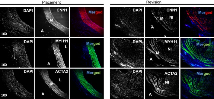
Figure 7.

Characterization of vascular smooth muscle cell (VSMC) differentiation program in human venous samples. Venous samples obtained from patients undergoing arteriovenous fistula (AVF) placement or at revision of a failed AVF were subjected to immunostaining for VSMC contractile proteins including CNN1, MYH11, and ACTA2. Representative images for each protein staining in the consecutive cross sections are shown. Strong staining of the three VSMC markers (CNN1, MYH11, and ACTA2) was seen in the medial layer (M) of the placement vessels. However, there was little to no signal in the adventitia (A). Strong staining of the 3 VSMC markers was also observed in the thickened medial layer of the failed revision AVF samples, but there was decreased intensity seen in the neointima (NI) of revision samples. L indicates the lumen of the vessel. The image is representative of 4 placement and 4 revision veins, each from different individuals (8 individuals in total). DAPI indicates 4′,6‐diamidino‐2‐phenylindole.
