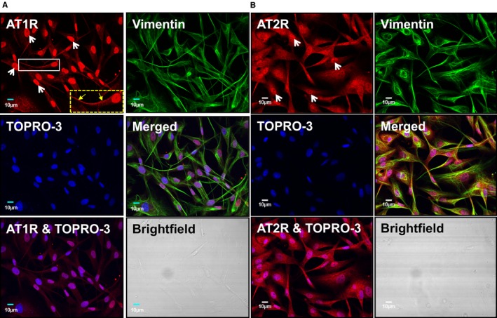
Figure 2.

AT1Rs and AT2Rs colocalize with TOPRO‐3 nucleic‐acid stain in canine atrial fibroblasts. A, Cultured atrial fibroblasts were permeabilized and then labeled with an AT1R antibody conjugated with Alexa Fluor 488 (red), anti‐vimentin conjugated with Alexa Fluor 555 (green), and TOPRO‐3 (blue). Merged images indicate the extent of colocalization. The dashed box at the lower right corner of the AT1R‐stained image shows an enlarged version of the fibroblast in the smaller white box. Linear membrane staining is indicated by the yellow arrows. B, Cultured atrial fibroblasts were permeabilized and then labeled with an AT2R antibody conjugated with Alexa Fluor 488 (red), anti‐vimentin conjugated with Alexa Fluor 555 (green), and TOPRO‐3 (blue). Merged images indicate the extent of colocalization. Brightfield images confirm the absence of cardiomyocytes from the fibroblast preparation. Similar observations were obtained from 5 different canine heart preparations for both AT1Rs and AT2Rs. AT1Rs indicates Ang‐II type 1 receptors.
