Figure 3.

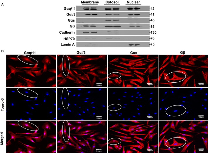
Presence of G protein subunits in fibroblast nuclei. A, Detection of Gαq/11, Gαi/3, Gαs, and Gβ in membrane, cytosolic, and nuclear fractions by immunoblot. Similar observations were obtained in fibroblasts isolated from each of 4 different dog hearts. The positions of molecular weight markers are indicated at the right (in kDa). B, Representative confocal images demonstrating the distribution of Gαq/11, Gαi/3, Gαs, and Gβ in permeabilized atrial fibroblasts. Superimposed confocal image showing the colocalization of G protein subunits with TOPRO‐3 double‐stranded DNA stain. The ovals outline illustrative cells. Gαq/11 and Gαi/3 protein staining (red) is clearly localized over the nucleus. Gαs and Gβ have much more diffuse distribution over the cell bodies, with some apparent perinuclear localization. Similar observations were obtained from 4 different canine heart preparations.
