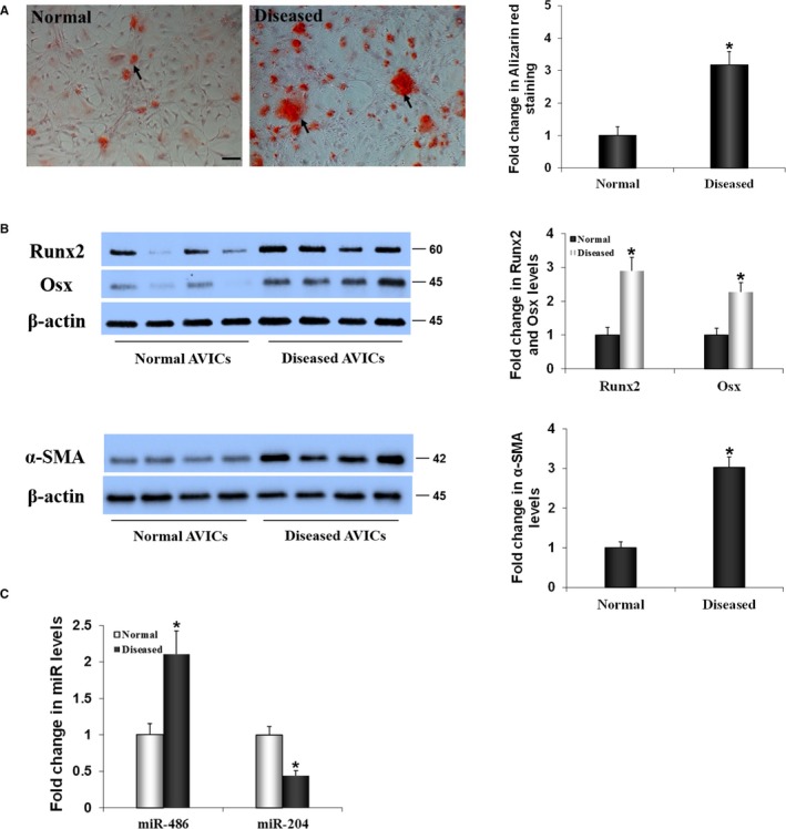
Figure 1.

The pro‐osteogenic phenotype of aortic valve interstitial cells (AVICs) of calcified valves is associated with altered expression of microRNAs miR‐486 and miR‐204. A, Representative images and spectrophotometric data show that AVICs from calcified human aortic valves exhibit greater calcium deposition when incubated in a conditioning medium (growth medium supplemented with 10 mmol/L β‐glycerophosphate, 10 nmol/L vitamin D3, 10 nmol/L dexamethasone, and 8 mmol/L CaCl2) for 14 days. Scale bar=200 μm. B, Representative images and spectrophotometric data show that AVIC isolates from calcified valves have higher levels of runt‐related transcription factor 2 (Runx2) and osterix (Osx), and α–smooth muscle actin (α‐SMA). C, Real‐time quantitative reverse transcriptase–polymerase chain reaction data in the bar graph show that miR‐486 levels are increased and miR‐204 levels are decreased in AVICs of calcified valves. Data are expressed as mean±SE. n=8 cell isolates from different valves in each group; independent samples t test and nonparametric Mann–Whitney U test; *P<0.05 vs normal.
