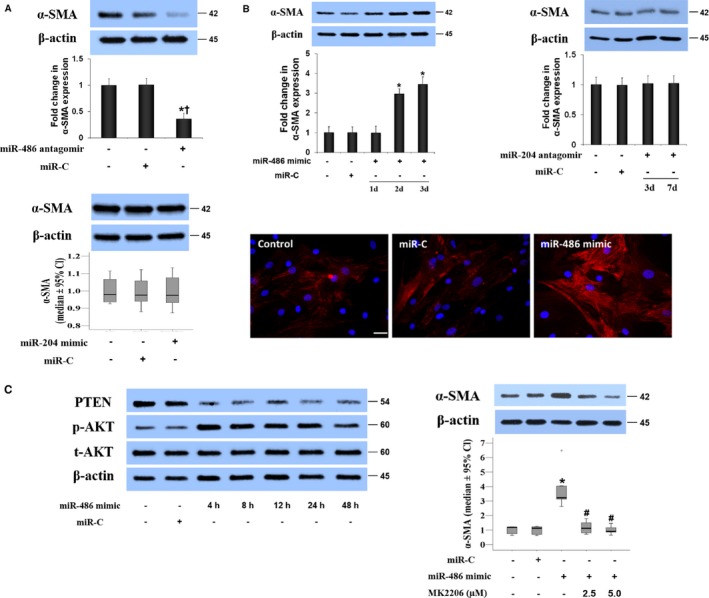
Figure 3.

MiR‐486 upregulates α–smooth muscle actin (α‐SMA) expression in aortic valve interstitial cells (AVICs) through the AKT signaling pathway. A, Representative immunoblots and quantitative analysis show that transfection of AVICs from calcified valves with miR‐486 antagomir reduces α‐SMA levels at day 3 after transfection, whereas transfection with miR‐204 mimic or control microRNA (miR‐C) has no effect. Data are expressed as mean±SE or median and 95% CI (α‐SMA levels in diseased cells treated with miR‐204 mimic or control miR). n=6 separate experiments using distinct cell isolates; ANOVA and nonparametric Kruskal–Wallis H test; *P<0.05 vs untreated AVICs of calcified valves; † P<0.05 vs AVICs of calcified valves plus miR‐C. B, AVICs of normal valves were transfected with miR‐486 mimic or control microRNA (miR‐C). Representative immunoblots of 5 separated experiments and quantitative analysis show that miR‐486 mimic increases α‐SMA levels in normal AVICs, whereas miR‐204 antagomir has no effect. Representative immunofluorescence images show the effect of miR‐486 mimic on α‐SMA expression. Scale bar=100 μm. C, Representative immunoblots show that miR‐486 mimic reduces cellular PTEN levels at 4 to 48 hours and induces AKT phosphorylation in normal AVICs. Inhibition of AKT with MK2206 abrogates the effects of miR‐486 mimic on α‐SMA expression. Data are expressed as mean±SE or median and 95% CI (α‐SMA levels). n=7 in separate experiments using distinct cell isolates; ANOVA and nonparametric Kruskal–Wallis H test; *P<0.05 vs control normal AVICs or normal AVICs plus miR‐C; # P<0.05 vs normal AVICs plus miR‐486 mimic.
