Fig. 1.
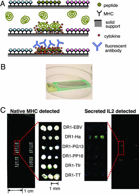
Artificial antigen-presentation chips. (A) Schematic representation of artificial antigen-presenting microarray technology. (Top) MHC–peptide complexes immobilized with different peptide antigens in distinct areas. Costimulatory and cytokine-capture antibodies can be coimmobilized (not shown). (Middle) T cells are incubated with the array. Only specific MHC–peptide complexes induce T cell responses. Cytokines are captured locally by immobilized anti-cytokine antibodies. (Bottom) Captured cytokines are detected by labeled antibodies in the locations where they were secreted, identifying the activating epitopes. (B) Photograph of a microarray with solution held in place by a hydrophobic barrier. (C) Microarray carrying various DR1–peptide complexes on a polystyrene slide, incubated with 106 murine hybridoma cells specific to DR1–Ha for 16 h and stained for native MHC with LB3.1-CY5 (Left) and for captured mouse IL-2 by using biotinylated α-mouse-IL-2 preincubated with SA-Alexa Fluor 555 (Right). The pattern was repeated in four areas.

