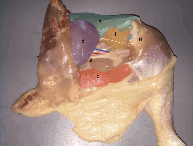
Fig. 4. Pedicle Dissection.

Femoro-cruralis (f) (yellow) is divided inferio-laterally and elevated superiorly to reveal the main femoral source vessels (blue and red lines over femoral vein and artery respectively). The adductor profundus (AP) muscle (d) (violet) is then freed by dividing its bony insertion (g) (white line) along with its septal attachment to the vastus lateralis (h) (green) superiorly. (c) Flexor cruris medialis, (e) muscle branch.
