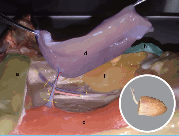
Fig. 6. Chicken thigh adductor profundus elevated.

The femoral source pedicle is further dissected distally to reach approximately 5–6 cm of pedicle length. The femoral source vessels are divided around the flap’s pedicle (yellow lines) to free the adductor profundus (d) muscle (violet) free flap. (a) Remnant abdominal muscl, (c) flexor cruris medialis, (f) femoro-cruralis, (h) vastus lateralis.
