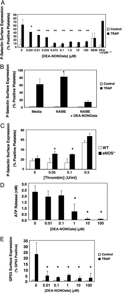
Fig. 1.
NO inhibition of platelet granule exocytosis. (A) Exogenous NO inhibits α-granule exocytosis: P-selectin. Human platelets were incubated with DEA-NONOate for 15 min and treated with 15 μM TRAP, and the amount of P-selectin on the surface was measured by FACS. (n = 6 ± SD, *, P = 0.05; **, P < 0.01 vs. TRAP without NO donor.) (B) Endogenous NO inhibits α-granule exocytosis. Human platelets were pretreated with 1 mM l-NAME for 15 min to inhibit endogenous NOS and treated with 15 μM TRAP, and the amount of P-selectin on the surface was measured by FACS. (n = 3–6 ± SD, *, P < 0.01 vs. 0 mM l-NAME.) (C) Endogenous NO inhibits α-granule exocytosis. Platelets from wild-type and eNOS-/- mice were treated with control or thrombin, and the amount of P-selectin on the surface was measured by FACS. (n = 3 ± SD, *, P < 0.01 vs. WT.) (D) NO inhibits dense-granule exocytosis. Human platelets were pretreated with DEA-NONOate for 15 min and treated with 15 μM TRAP, and the amount of released ATP was measured by chemiluminescence. (n = 3 ± SD, *, P < 0.01 vs. 0 μM.) (E) NO inhibits lysosomal granule release. Human platelets were pretreated with increasing amounts of DEA-NONOate for 15 min and treated with 15 μM TRAP, and the amount of gp53 (CD63) on the surface was measured by FACS. (n = 3 ± SD, *, P = < 0.01 vs. 0 μM.)

