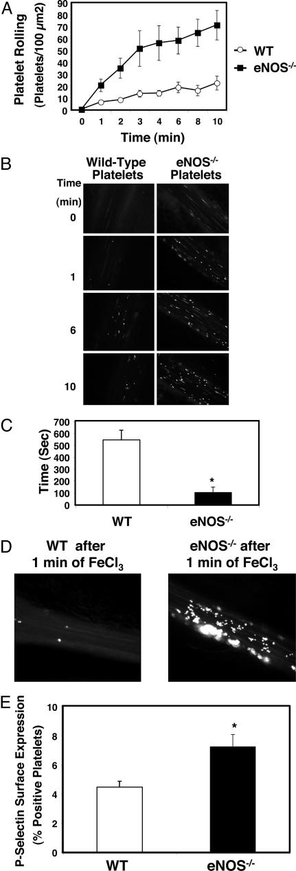
Fig. 5.
NO inhibits platelet adherence and aggregation in vivo. (A) Platelet rolling. Wild-type mice were anesthetized and injected with calcein-AM-labeled platelets from wild-type (WT) or eNOS-/- mice. The mesentery was externalized, and venules 120–150 μm in diameter were treated with 250 mM FeCl3. Platelet rolling on venules was imaged with a digital fluorescent camera (n = 7–8 ± SEM). (B) Digital fluorescence images of platelet rolling. Wild-type mice were anesthetized and injected with calcein-AM-labeled platelets from wild-type or eNOS-/- mice. The mesentery was externalized, and venules 120–150 μm in diameter were treated with 250 mM FeCl3. Platelet rolling on venules was imaged with a digital fluorescent camera 0, 1, 6, and 10 min after FeCl3 treatment. (Representative images from n = 7–8.) (C) Thrombosis. Mice were injected with labeled platelets as above. The mesentery was externalized, and arterioles 50–80 μm in diameter were treated with 500 mM FeCl3. The time to formation of the first thrombus >10 μm in size was recorded (n = 5 ± SD). (D) Digital fluorescent images of thrombosis. Wild-type and eNOS-/- mice were injected with labeled wild-type platelets, arterioles were treated with FeCl3 as above, and a digital fluorescent camera was used to visualize formation of the first thrombus >10 μm in size. (E) Exocytosis in shed platelets. The distal 3 mm of wild-type and eNOS-/- mice tails were amputated, and shed blood was collected. Platelets were analyzed for surface P-selectin expression by FACS. Shed platelets from eNOS-/- have increased exocytosis as measured by P-selectin expression compared with WT mice. (n = 3 ± SD, *, P < 0.002 for WT vs. eNOS-/-.)

