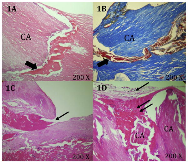
Fig. 2. Characteristics of the tissues surrounding plaque fissures.
(A and B) H&E and Mallory’s stained plaque showing hemorrhage (arrows) dissecting the tissue planes between matrix and calcifications. (C) H&E stained plaque with fissure containing a small luminal fibrin thrombus (arrow). (D) Loose matrix (arrow) and thrombus (double arrow) covering the fissure at the luminal surface.

