Fig. 3.
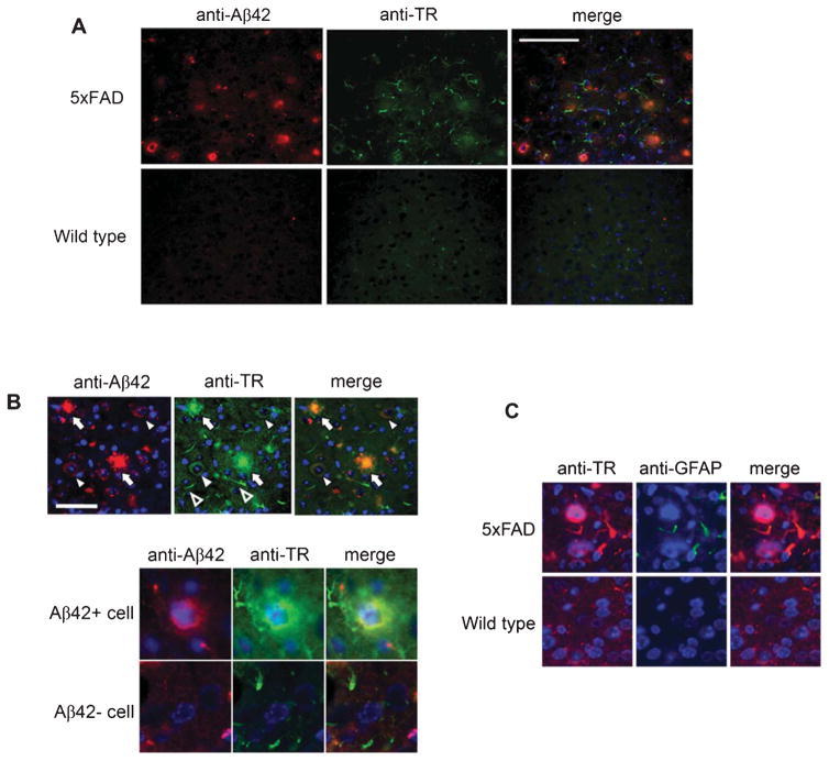
TR is associated with amyloid plaques and intraneuronal Aβ, and is expressed in reactive astrocytes in 5xFAD mice. A) Cortical sections of 5xFAD and age-matched wild-type littermate mice were co-immunostained with anti-Aβ42 and anti-TR. Scale bar = 100 μm. B) Three TR-immunoreactive structures are indicated: Arrows: amyloid plaques; Filled arrowheads: neurons with intraneuronal Aβ; and Empty arrowheads: curvilinear profiles. Scale bar = 50 μm. The lower panel shows magnified images of a neuron with intraneuronal Aβ in comparison to one without. C) Co-immunostaining with anti-TR and anti-GFAP showed that the TR-positive curvilinear profiles were processes of reactive astrocytes in the neighborhood amyloid plaques.

