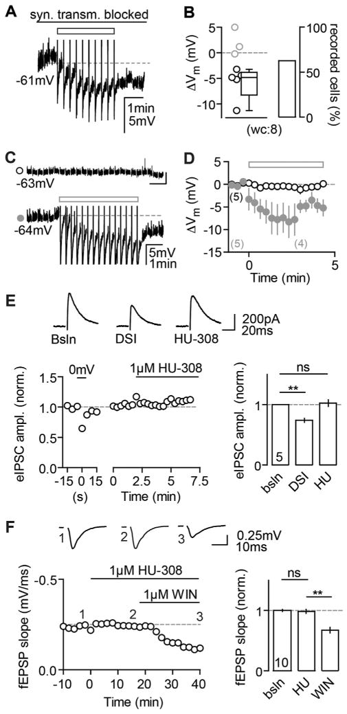
Figure 7. Comparison of CB2R- versus Presynaptic CBR-Mediated Effects.
(A and B) The continuous block of glutamatergic (20 μM NBQX, 50 μM D-AP5) as well as GABAergic (1 μM Gabazine, 1 μM CGP) transmission does not abolish the AP-induced hyperpolarization. (A) Example wc recording of a reactive CA3 PC in response to the AP train (rectangle) is shown. (B) The ΔVm of each recorded cell (circles) and the median and 25th and 75th percentiles of the average ΔVm of all reactive cells are shown for n(N) = 8(2) experiments (−4.8, −8.3, and −3.8 mV; left). The percentage of reactive cells is 62.5% (right).
(C) Dual pp recording of 2 CA3 PCs. AP firing in a control cell (lower trace) elicits a hyperpolarization in this, but not in the other cell (upper trace). The AHPs of the control cell are clipped for display purposes.
(D) In 5/5 recordings (N = 5), the control cell hyperpolarized in response to the AP trains (filled gray circles, ΔVm: −6.0 ± 1.5 mV), whereas the unstimulated cell did not (open black circles, ΔVm: 0.02 ± 0.6 mV).
(E and F) CB2R agonists cannot mimic CB1R-mediated depression of synaptic transmission. (E) HU has no effect on DSI-positive eIPSCs. Left: example of a CA3 PC recorded in wc configuration is shown. Depolarization of the neuron results in a transient reduction of eIPSC amplitude, whereas bath application of HU does not. Right: mean normalized eIPSC amplitudes of n(N) = 5(4) experiments for DSI (0.7 ± 0.03) and HU application (1 ± 0.05) in comparison to baseline (paired t test, p = 0.0016 and p = 0.67) are shown. (F) WIN, but not HU, suppresses evoked field responses in CA3. Left: exemplary fEPSP recording with HU and WIN bath application is shown. Right: mean normalized fEPSP slopes for HU (1 ± 0.03) and WIN (0.7 ± 0.05) in comparison to baseline (paired t test, p = 0.33 and p < 0.001) are shown.

