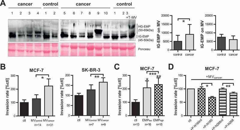
Figure 5.

Highly-glycosylated EMMPRIN is enriched on cancer-patient-derived MV and mediates tumour invasion. (a) Western blot analysis of EMMPRIN glycoforms on MV derived from cancer or control patients. Ponceau staining is depicted as loading control. In the last control sample (first lane on the right), in vitro T-MV derived from MCF-7 cells were added to demonstrate the increase of HG-EMMPRIN in the presence of tumour-derived MV. HG-EMP = highly-glycosylated EMMPRIN, IG-EMP = intermediately-glycosylated EMMPRIN. Western blot images depict one representative of five independent experiments. Histograms represent mean signal intensities ± SD of HG- and IG-EMP in the MV analysed by western blot. *p = 0.035 with Wilcoxon rank sum test. (b) Boyden chamber assays of MCF-7 and SK-BR-3 cells stimulated for 96 h with 1 µg/ml MV derived from cancer or control patients (n = 14 for MCF-7, n = 4 for SK-BR-3, mean±SD, *p = 3.288e-17, **p = 7.833e-10 with two-sided t test). Invasion was calculated compared to unstimulated cells (ctl). (c) Invasiveness of MCF-7 cells either unstimulated (ctl) or in the presence of 1µg/ml MV derived from cancer patients with a high (>35%) or low (<35%) percentage of EMMPRIN+ MV in blood (n = 14, mean±SD, *p = 8.652e-17, **p = 4.379e-23, ***p = 0.031 with two-sided t test). (d) Boyden chambers: MCF-7 cells were pre-incubated for 2 h with or without (=ctl) specific blocking peptides directed towards N160 (P-N160) and N268 (P-N268) or random control peptides (P-N160rd and P-N268rd) and then stimulated with 1 µg/ml cancer-patient-derived MV (n = 10, mean±SD, *p = 7.722e-05, **p = 1.075e-03 with two-sided t test).
