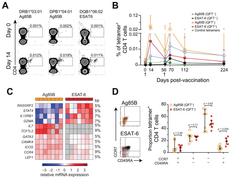
Figure 4. Transcriptomic profiles show that human ESAT-6-specific CD4 T cells are more differentiated cells than Ag85B-specific cells during latent Mtb infection.
(A) Ag85B and ESAT-6 HLA class II tetramer staining of CD4 T cells from H1:IC31 vaccinated adolescents (day 14). HLA allele-matched tetramers bearing irrelevant peptide antigens were used as controls. Numbers represent the frequencies of tetramer+ CD4 T cells.
(B) Median (error bars denote IQR) frequencies of tetramer+ CD4 T cells in QFT− or QFT+ adolescents who received two vaccinations of H1:IC31. The arrows indicate vaccine administration time-points.
(C) Supervised heat map of 11 mRNA transcripts differentially expressed (Mann-Whitney U test p < 0.05 and False discovery rate (FDR) < 10%) between tetramer-sorted Ag85B-specific and ESAT-6-specific CD4 T cells from QFT+ adolescents, 14 days after H1:IC31 vaccination. Undetected mRNAs are depicted in grey.
(D) CCR7 and CD45RA expression by total CD4 T cells (grey background), Ag85B-specific (orange), or ESAT-6-specific (red) tetramer+ CD4 T cells. FDRs were calculated using the Benjamini-Hochberg method and p-values were calculated using the Mann-Whitney U test. See also Figure S5.

