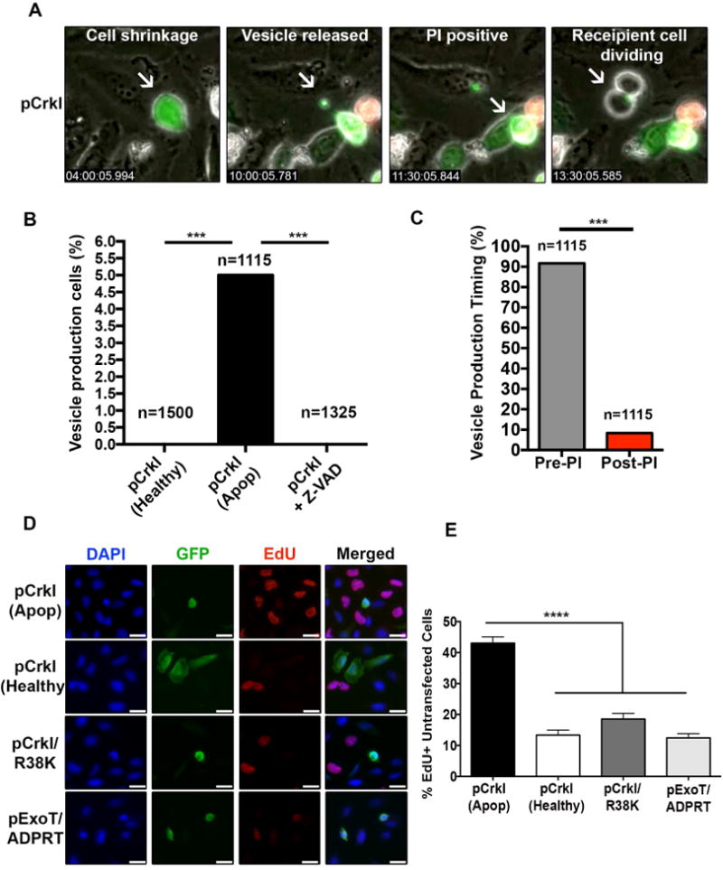
Fig. 1. Apoptotic cells produce and release CrkI-containing vesicles, which appear to be capable of inducing proliferation in bystander cells.

HeLa cells were transfected with CrkI-GFP in the presence or absence of Z-VAD and followed by IF time-lapse videomicroscopy. (A) Selected movie frames of a CrkI-transfected apoptotic cell are shown. This cell releases a CrkI-containing microvesicle, before it becomes PI positive, and the bystander cell initiates mitosis after contacting this vesicle. (B) The percentages of non-apoptotic, apoptotic, and Z-VAD-treated, CrkI-GFP transfected cells producing vesicle is shown. (C) The ability of CrkI-GFP transfected apoptotic cells to produce and release vesicles prior or post PI uptake was assessed by IF time-lapse microscopy and the tabulated results are shown. (For B & C, n≥1115, *** p<0.001, χ2 analysis). (D-E) Cells were transiently transfected with CrkI-GFP, CrkI/R38K-GFP, or ExoT/ADPRT-GFP expression vectors. 17h after transfection, cells were treated with EdU for 2h, fixed by 10% TCA, and analyzed for EdU incorporation in untransfected cells surrounding transfected apoptotic cells (identified by cell shrinkage/rounding) or healthy (identified by spread-out morphology). Representative images are shown in (D) and the tabulated data are shown in (E) (n≥3 independent experiments, 10 random fields/experiment, **** p<0.0001, One-way ANOVA).
