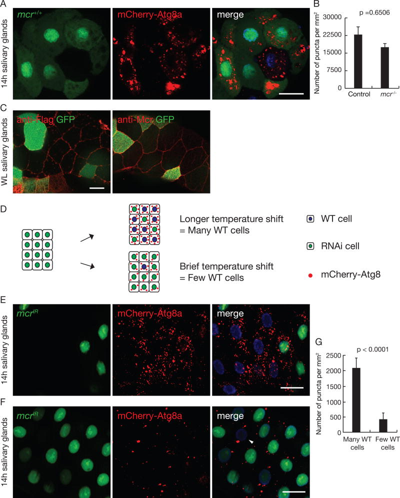
Figure 4. Mcr regulates autophagy in a cell non-autonomous manner in salivary glands.
(A) Salivary glands dissected 14h after puparium formation containing a loss-of-function mcrEY07421 mutant cell clone (lacking GFP), imaged for mCherry-Atg8a (red), GFP (green) and Hoechst (blue). Scale bars, 50 µm.
(B) Quantification of data from (A). Error bars, mean ± SEM; n = 24 (control), n = 19 (mcr −/−). Statistical significance: Student’s t-test.
(C) Salivary glands expressing Mcr-flag specifically in GFP-marked cells at wandering third instar larval stage were dissected and stained with antibodies against Flag (left) and Mcr (right). Scale bars, 20 µm.
(D) Strategy for Gal80 flip-in induction of salivary glands with variable numbers of mcrIR expressing salivary gland cells.
(E and F) Salivary glands dissected 14h after puparium formation and imaged for mCherry-Atg8a puncta (red) after varying the numbers of mcrIR-expressing cells (green, GFP-positive). Following temperature shift, Gal80 is expressed in a subset of cells, repressing Gal4 activation of UAS-mcrIR. There are more mCherry-Atg8a puncta in salivary glands with many wild-type cells (lacking green GFP) (E), compared to glands with one or two wild-type cells (arrow head, F). Scale bars, 50 µm.
(G) Quantification of data from (E) and (F). Error bars, mean ± SEM; n = 12 (many WT cells), n = 12 (few WT cells). Statistical significance: Student’s t-test.
See also Figure S3.

