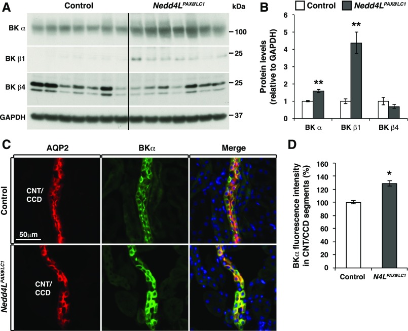
Figure 7.
BK channels are upregulated in CNT/CCD principal cells of Nedd4LPax8/LC1 mice. (A and B) Western blot analysis of BK in control and Nedd4LPax8/LC1 mice after 2 weeks of LKD, and protein quantification revealing a significant increase in BKα and BKβ1 expression (seven mice of each genotype). (C) Costaining of BKα (green) and AQP2 (red) in control and Nedd4LPax8/LC1 mice. (D) Quantification of BKα fluorescence intensity in the CNT/CCD segments. BKα labeling was significantly increased in the principal cells of mutant CNT/CCD segments. *P<0.05; **P<0.01.

