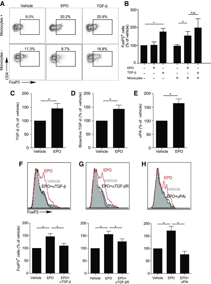
Figure 1.
EPO promotes in vitro human Treg induction. (A and B) Enriched naïve human CD4+ T cells were cultured for 5 days in the presence (top) or absence (bottom) of CD14+ monocytes, anti-CD3 (1 μl/ml), IL-2 (100 IU/ml), and EPO (1000 IU/ml) or vehicle control. Naïve CD4+ T cells cultured in the presence of anti–CD3- and anti–CD28-coated beads (25 μl/106 cells), IL-2, and TGFβ (5 ng/ml) were used as positive control (right panel in each row). Representative flow plots for FOXP3 expression gated on CD4+CD25+ T cells (A) and summarized normalized quantification (B) (see Statistical Analyses) of 15–22 experiments using different donors. (C–E) Human monocytes were cultured in serum-free media with EPO (1000 IU/ml) or vehicle control for 2 days and culture supernatants tested for total TGFβ by ELISA (C), bioactive TGFβ using SMAD3-SMAD4 reporter cells (D), and uPA protein (ELISA) (E). (F–H) Enriched human naïve CD4+ T cells were cultured for 5 days in the presence of monocytes, anti-CD3 mAb (1 μl/ml), IL-2 (100 IU/ml), and EPO (1000 IU/ml) or vehicle control in serum-free media. (F–H) Treg induction experiments as performed in (A and B), in which (F) anti-TGFβ antibody (10 μg/ml), (G) anti-TGFβ receptor neutralizing antibody (10 μg/ml), or (H) uPA chemical inhibitor (100 μM) were added to the cultures and compared with relative isotype or vehicle controls. Top panels show representative flow plots for FOXP3 expression gated on CD4+CD25+ T cells, bottom panels show summarized quantification of three independent experiments from nine different donors. *P<0.05 (paired t test).

