Figure 5.
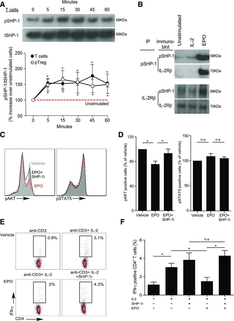
EPO/EPO-R binding inhibits IL-2Rβ signaling in Tconv via SHP-1 molecular crosstalk. (A) Enriched human total T cells and CD4+CD25+ Treg were stimulated with EPO (1000 IU/ml) for the indicated times and the lysates were immunoblotted for pSHP-1 and tSHP-1. The top panel shows a representative gel using total T cells and the bottom panel shows ratios of densitometry measurements of pSHP-1 to tSHP-1 band intensity for T cells and Treg as indicated. *P<0.05 versus unstimulated cells (two-way ANOVA). (B) Immunoprecipitation experiments. Cell lysates were produced from human resting total T cells or from total T cells activated with 100 IU/ml IL-2 (labeled IL-2) or IL-2 plus 1000 IU/ml EPO (labeled EPO) for 60 minutes. The cell lysates were immunoprecipitated with either anti–pSHP-1 (B) (top) or anti–IL-2Rβ (B) (bottom), and blotted with anti–pSHP-1 or anti–IL-2Rβ as indicated. Representative of three individual experiments. (C and D) Total T cells were activated with anti–CD3- and anti–CD28-coated beads (25 μl/106 cells), IL-2 (100 IU/ml), and EPO (1000 IU/ml), EPO plus SHP-1 inhibitor (NCS-87877; 175 μg/ml), or vehicle control for 30 minutes. pAKT and pSTAT5 were assayed by flow cytometry. (C) Representative plots and (D) summarized data quantification of six to nine experiments. *P<0.05 (paired t test). (E and F) Human PBMC were activated with anti-CD3 (1 μg/ml) with or without IL-2 (100 IU/ml), with or without EPO (1000 IU/ml), and with or without SHP-1 inhibitor (NCS-87877; 37.5 μg/ml) for 24 hours. (E) Representative flow plots (gated on CD4+ T cells) and (F) summarized quantification of intracellular IFNγ expression from four independent experiments with different donors. *P<0.05.

