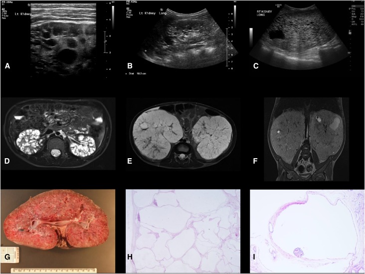
Figure 2.
Renal imaging and histology in children with HIPKD. (A) Ultrasound images of the kidney from patient 2.1, left kidney, age 11 years; (B) patient 2.2, right kidney, age 4 years; and (C) patient 6.1, right kidney, age 2 years. Note the presence of cysts of various sizes. Kidney length was >95th percentile for age in all patients. (D) Axial MRI image of the kidney from patient 2.1, age 11 years; (E and F) axial and coronal MRI images (without contrast) from patient 6.1, age 2 years. Note the massively enlarged kidneys with cysts of various sizes. (G) Macroscopic appearance of the nephrectomy specimen from patient 6.1, age 2 years, removed at the time of transplant. Note the large kidney size (20 cm longitudinal, normal <7.7 cm) and numerous macroscopic cysts. (H and I) Histology of the same kidney demonstrating multiple cysts lined by simple attenuated epithelium, with glomeruli noted in some (glomerulocystic disease; hematoxylin and eosin; original magnifications, ×20 and ×100, respectively). Dist, distance; Lt, left; Rt, right.

