
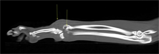
Figure 2.
Sagittal multiplanar reformatted image of the left carpus of case 2. There is marked soft tissue swelling associated with the antebrachiocarpal joint. Osteolysis of the distal radius is also identified (arrows)
Official websites use .gov
A
.gov website belongs to an official
government organization in the United States.
Secure .gov websites use HTTPS
A lock (
) or https:// means you've safely
connected to the .gov website. Share sensitive
information only on official, secure websites.
Sagittal multiplanar reformatted image of the left carpus of case 2. There is marked soft tissue swelling associated with the antebrachiocarpal joint. Osteolysis of the distal radius is also identified (arrows)