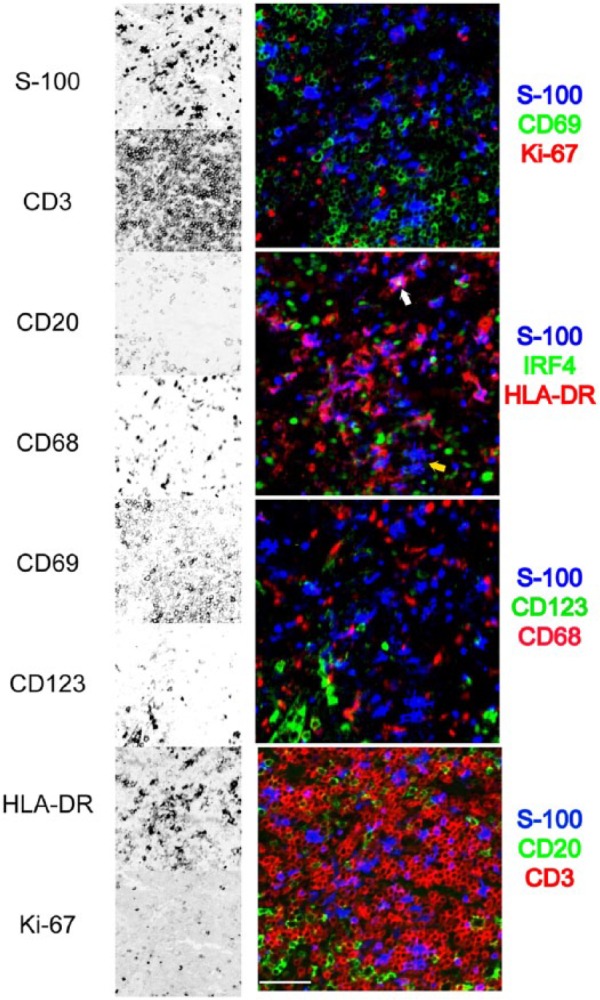
Figure 8.

Detail of a multiplexed interfollicular tonsil area. Eight stains out of a 32-antibody multiplex are selected from a tonsil interfollicular area and shown as single stains (inverted gray scale) on the left, or combined into four three-color RGB composites. The area contains numerous S-100+ dendritic cells, HLA-DR+ with exceptions (yellow arrow), occasionally IRF4+ (white arrow), negative for Ki-67, CD69, CD68, and CD123. The dendritic cells are located in a CD3+ T-cell area, without contact with CD20+ B cells. GnHCl stripping method. Scale bar = 100 µm. Abbreviations: RGB, red green blue additive color; GnHCl, guanidinium hydrochloride.
