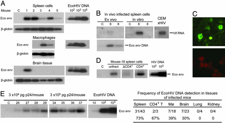
Fig. 2.
Conventional mice are susceptible to EcoHIV infection. Mice were inoculated once with EcoHIV and numbered or mock-inoculated and maintained in parallel, labeled as C. (A) Six weeks after infection, mice were killed and DNA from spleen, brain, and macrophages was subjected to PCR, amplifying a region in the chimeric envelope gene unique to EcoHIV; input was standardized by amplification of β-globin. At right is a standard curve of amplification of the indicated number of copies of plasmid DNA. (B) After infection (3 mo), mice were killed, spleens were collected, and either DNA or RNA was isolated or cells were cultured. (Upper) Amplification of singly spliced Vif mRNA in fresh cells or cells after 3-d culture. HIV-1-infected human CEM cells serve as positive controls. (Lower) Amplification of EcoHIV DNA in the same samples. (C) Spleen cells harvested 6 wk after EcoHIV infection were serially cocultured three times with uninfected cells and stained for HIV-1 antigens. (Upper) Infected cells. (Lower) Uninfected cells. (D) Spleen cells harvested 3 mo after infection were subjected to magnetic bead fractionation for CD4-bearing cells before DNA PCR; unfract, unfractionated spleen cells; ΔCD4+, CD4-negative spleen cells. (E) Mice were inoculated once with EcoHIV at the indicated doses, and spleen cells were harvested 6 wk after infection and subjected to DNA PCR. The table at the right summarizes six independent experiments testing the indicated tissues for EcoHIV DNA.

