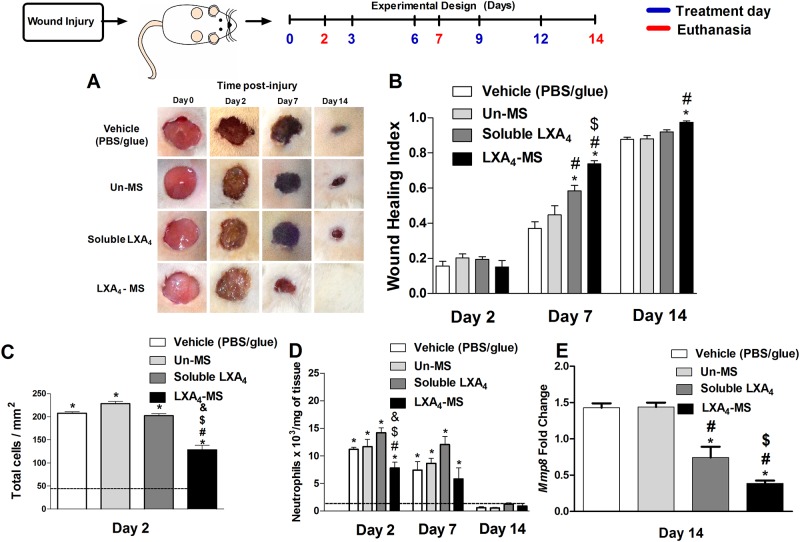
Fig 2. Topical application of LXA4-MS to skin ulcers accelerated wound closure and attenuated neutrophil chemotaxis.
(A) Representative images of 1.5 cm dorsal wounds were collected on days 0, 2, 7, and 14 for the following groups: control (vehicle—PBS/glue), Un-MS, soluble LXA4, and LXA4-MS. (B) Wound healing index values for the groups outlined in (A). Index values range from 0 to 1, where a value of 0 indicates the original wound, and a value of 1 represents a completely closed wound. Values are means ± SEM (n = 10 ulcers/group). One-way ANOVA was done to determine statistical significance (p < 0.05), which is as follows: *, soluble LXA4 or LXA4-MS vs. vehicle (PBS/glue); #, LXA4-MS or soluble LXA4 vs. Un-MS; and $, LXA4-MS vs. soluble LXA4. (C) ImageJ software was used to count inflammatory cells on day 2 in at least 12 random optical 400× fields per group. (D) Neutrophil accumulation (represented as bars) as measured by MPO. Values are means ± SEM (n = 5 wounds/group). One-way ANOVA was done to determine statistical significance (p < 0.05), which is indicated as follows: *, demonstrated significant increase compared to normal tissue (dashed line); #, soluble LXA4 or LXA4-MS vs. Vehicle (PBS/glue); $, LXA4-MS or soluble LXA4 vs. Un-MS; and &, LXA4-MS vs. soluble LXA4. (E) qRT-PCR was performed to assess MMP8 mRNA transcript abundance in skin ulcers collected on days 2, 7, and 14 from the vehicle (PBS/glue), Un-MS, soluble LXA4, and LXA4-MS groups. Data represent means ± SEM (n = 5 ulcers/group). One-way ANOVA was done to determine statistical significance (p < 0.05) and indicated as follows: *, soluble LXA4 or LXA4-MS vs. Vehicle (PBS/glue); #, LXA4-MS or soluble LXA4 vs. Un-MS; and $, LXA4-MS vs. soluble LXA4.

