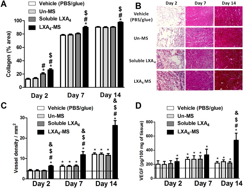
Fig 4. LXA4-MS increased collagen deposition and angiogenesis and affected VEGF production.
(A) Collagen deposition was measured using the ImageJ software with the Color Deconvolution plug-in, which measured densitometry in at least 12 random 400× fields in all groups, at days 2, 7 and 14. Percentages of collagen deposition are represented as means ± SEM (n = 6 ulcers/group). One-way ANOVA was used to determine statistical significance (p < 0.05) and is indicated as follows: *, soluble LXA4 or LXA4-MS vs. Vehicle (PBS/glue); #, LXA4-MS or soluble LXA4 vs. Un-MS; and $, LXA4-MS vs. soluble LXA4. (B) Photomicrographs of wounds stained with Picro Sirius Red (200×) show collagen deposition at days 2, 7, and 14. (C) Histogram showing quantitative analysis of vascular density using the ImageJ software with the Cell Counter plug-in on 200× images. One-way ANOVA was performed to determine statistical significance (p < 0.05), which is indicated as follows: *, significant VEGF increase as compared to normal tissue (dashed line); #, soluble LXA4 or LXA4-MS vs. Vehicle (PBS/glue); $, LXA4-MS or soluble LXA4 vs. Un-MS; and &, LXA4-MS vs. soluble LXA4. (D) VEGF was quantified in all groups at days 2, 7 and 14 (represented as bars) in wounds via ELISAs as proxy to blood vessel density. Values are means ± SEM (n = 6 wounds/group). One-way ANOVA was performed to determine statistical significance (p< 0.05), which is indicated as follows: *, significant VEGF increase as compared to normal tissue (dashed line); #, soluble LXA4 or LXA4-MS vs. Vehicle (PBS/glue); $, LXA4-MS or soluble LXA4 vs. Un-MS; and &, LXA4-MS vs. soluble LXA4.

