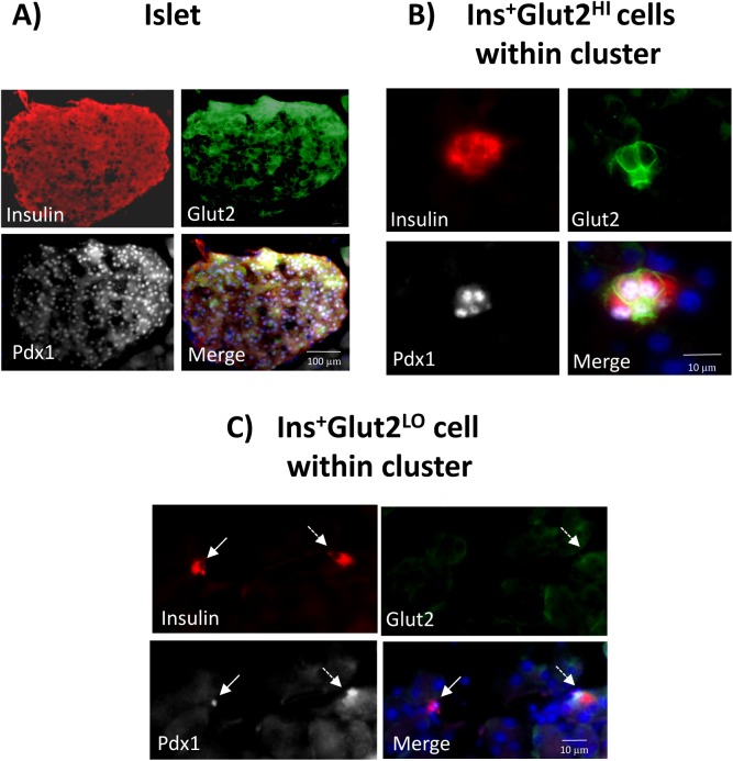
Fig 5. Co-localization of Pdx1 (white) with insulin (red) or Glut2 (green) in mouse pancreas at gestational day (GD) 9.
Nuclei were counter-stained with DAPI (blue). An islet of Langerhans is shown in A, and small β-cell clusters in B and C. Panel B shows a cluster containing Ins+Glut2HI cells demonstrating the presence of nuclear Pdx1together with cytoplasmic insulin and Glut2 associated with the plasma membranes. Panel C shows an Ins+Glut2LO cell (solid arrow) within a small cluster containing nuclear Pdx1as well as cytoplasmic insulin, but lacking detectable Glut2. To the right is shown an Ins+ cell with detectable Glut2 and nuclear-localized Pdx1(dashed arrow).

